Overview
In the field of physical therapy, understanding imaging interpretations is crucial for accurate diagnosis and treatment planning. This module aims to equip physical therapists with the knowledge and skills necessary to interpret X-ray images effectively, focusing on common themes PTs will encounter in the field, such as juvenile hip conditions, cervical spinal pathology, common anatomic variants as well as discussion topics on atypical foot/ankle presentation(s). Additionally, the module will explore the Pittsburgh and Ottawa rules, providing a comparative analysis to aid in clinical decision-making.
Educational methods:
- Lectures with visual aids (radiographs, diagrams, etc.)
- Case studies and clinical scenarios
- Group discussions and peer learning
- Online resources for further reading and self-assessment
This module has two assignments. One will be a discussion forum where you can make a recommendation based on the radiograph of a patient complaining of foot pain and then collaborate with your classmates. The second will involve assessing and evaluating different radiograph images and delving deeper into the Pittsburgh and Ottawa rules. For each assignment, you will research other resources and support your responses in both the discussion forum and your assignment with at least one additional reference.
By mastering the interpretation of X-ray images, such as identifying juvenile hip conditions and cervical spine abnormalities, and understanding the application of Pittsburgh and Ottawa rules, physical therapists can enhance their clinical skills and contribute to more accurate diagnoses and effective treatment plans. This module provides a comprehensive foundation for integrating imaging interpretation into physical therapy practice, ultimately improving patient outcomes and healthcare delivery.
Module objectives
By the end of this module, students will be able to:
- Identify radiograph views of different parts of the body.
- Describe the implications for physical therapy treatment for radiographs.
- Compare and contrast the Pittsburgh and Ottawa rules for ordering radiographs.
- Assess fractures and other abnormal pathologies on radiographs and describe the rehab implications.
- Perform an ABCS & D evaluation on a radiograph.
Readings
Course textbook
McKinnis, L. N. (2021). Fundamentals of musculoskeletal imaging (5th ed.). F. A. Davis Company.
- Chapter 2: Radiologic Evaluation, Search Patterns, and Diagnosis
The textbook is offered as an electronic book through the University's library, free with unlimited access.
Articles
Implementation of Ottawa Ankle Rules in University Hospital Emergency Room: Pilot Study. (2023)
This study discusses how implementing a protocol based on Ottawa Ankle Rules reduced X-rays, costs, radiation exposure, and improved efficiency in treating ankle sprains.
Validation of the Ottawa ankle rules: Strategies for increasing specificity. (2021).
This study discusses using Ottawa Ankle Rules to reduce unnecessary X-rays for ankle injuries.
Can the Ottawa and Pittsburgh rules reduce requests for radiography in patients referred to acute knee clinics? (2012).
This study investigates whether Ottawa and Pittsburgh rules can be used to avoid unnecessary X-rays for knee injuries.
Media presentations
Introduction to Radiograph Interpretations
This study investigates whether Ottawa and Pittsburgh rules can be used to avoid unnecessary X-rays for knee injuries.
Shoulder Evaluations with Dr. Colin Rigney
Assignments
Assignment 1
Foot case discussion board
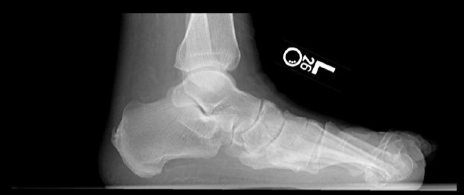
The radiograph above is a lateral view of a 58-year-old female with a history of long-standing diabetes. She has a history of several foot surgeries and is now complaining of general foot pain and difficulty walking. Clinical Decision Rules (CDR) are tools for helping clinicians make diagnostic and therapeutic decisions during initial patient evaluations. They provide a universal language and set of principles to avoid imaging overuse. The Ottawa Ankle Rules are one example of a CDR.
Discuss the following points in your original post. Read your classmate’s viewpoints on the case and share your thoughts about their recommendations. As you work on this assignment, reflect on your experience as a clinician and how your new knowledge can benefit your practice.
- Was it appropriate for the physician to order X-rays on this patient based on the Ottawa Ankle Rules? Please discuss and support your answer.
- Review the radiograph and discuss a pathological finding that might be present.
- Discuss what type of gait deficiencies this patient may have and what could contribute to her complaints.
- What other ankle view might be necessary to assess this patient’s foot properly? Support your answer.
Assignment 2
Part 1: Cervical spine images evaluation
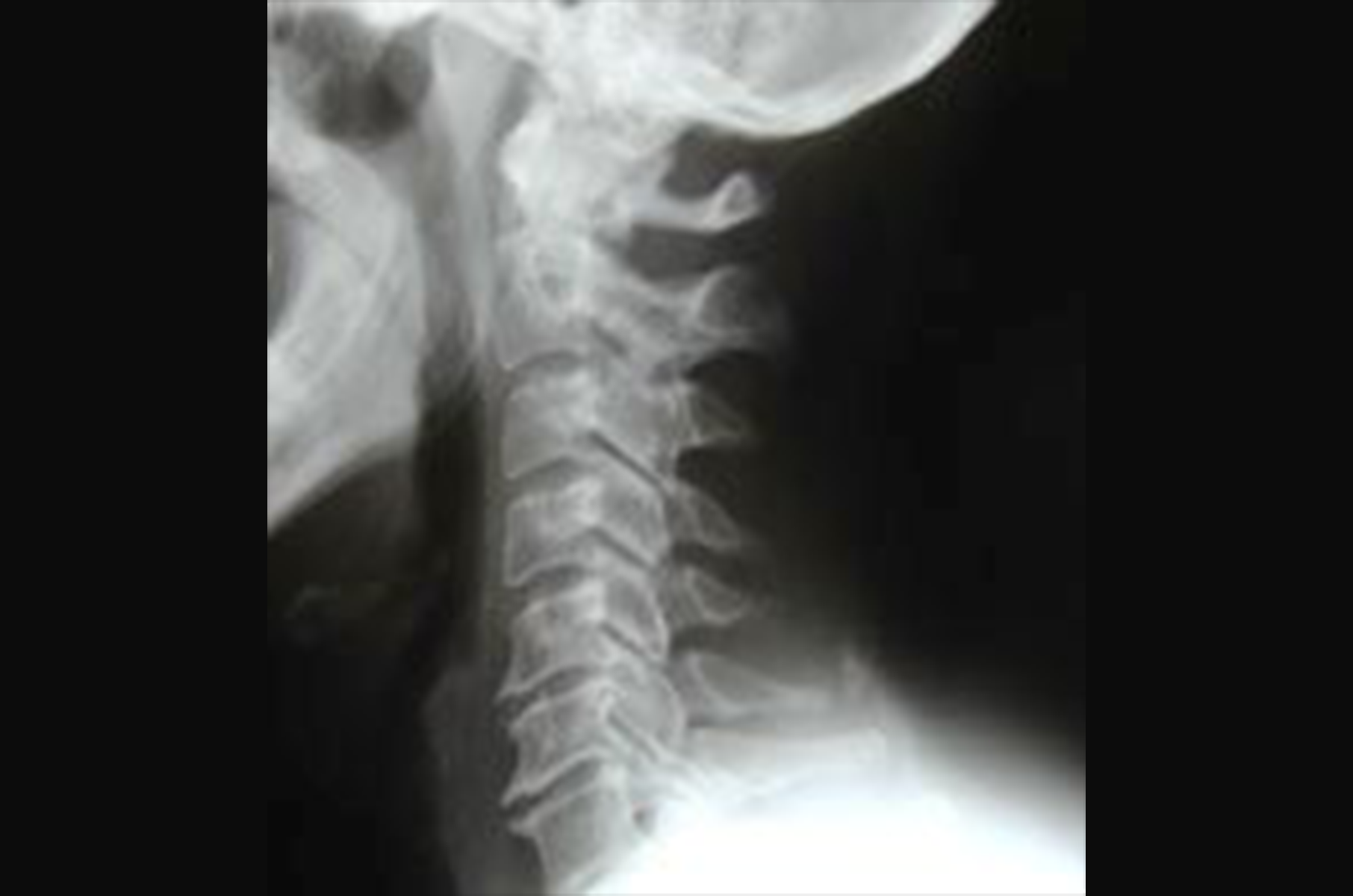
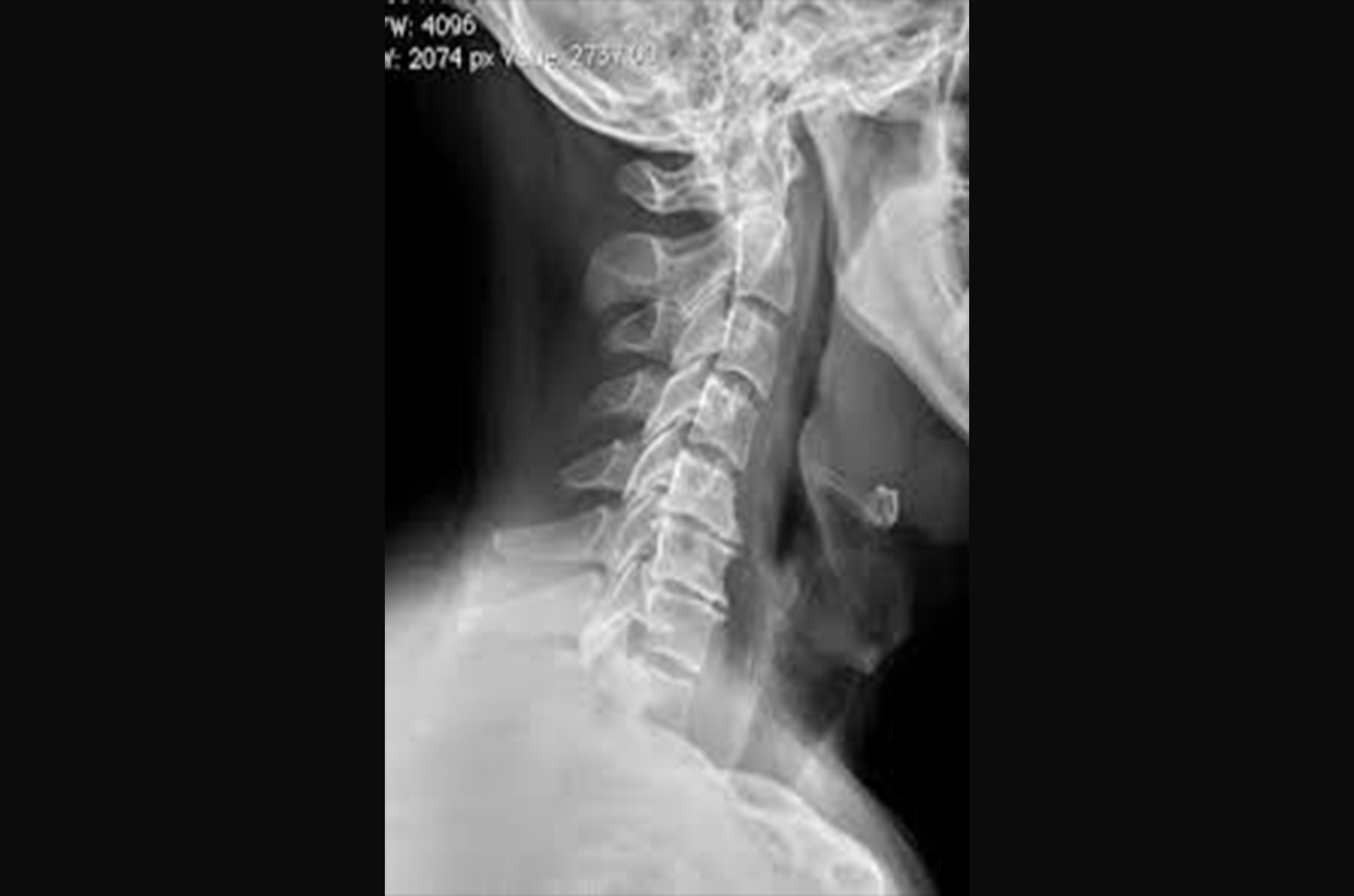
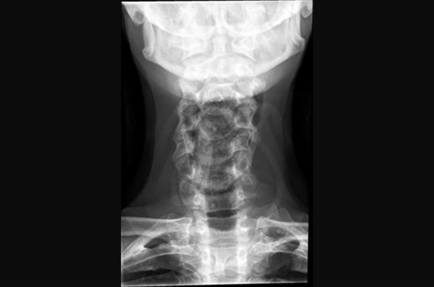
- X-ray 1-3: Identify the three views depicted in cervical spine images. Which cervical view is not depicted in this series?
- X-ray 1: Perform an ABCs & D evaluation. Provide a thorough discussion about the whole image. Use X-ray images #2 and #3 to support and identify any abnormalities in X-ray image #1. Discuss some of the clinical presentations this patient may exhibit.
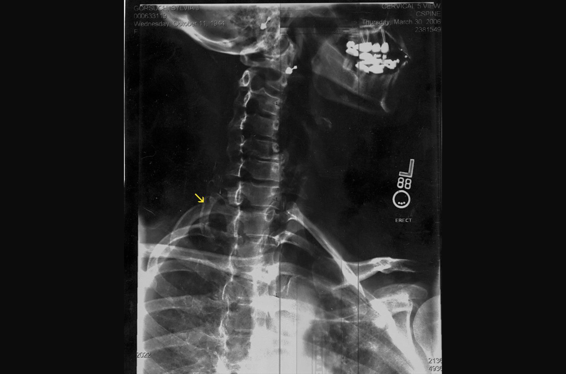
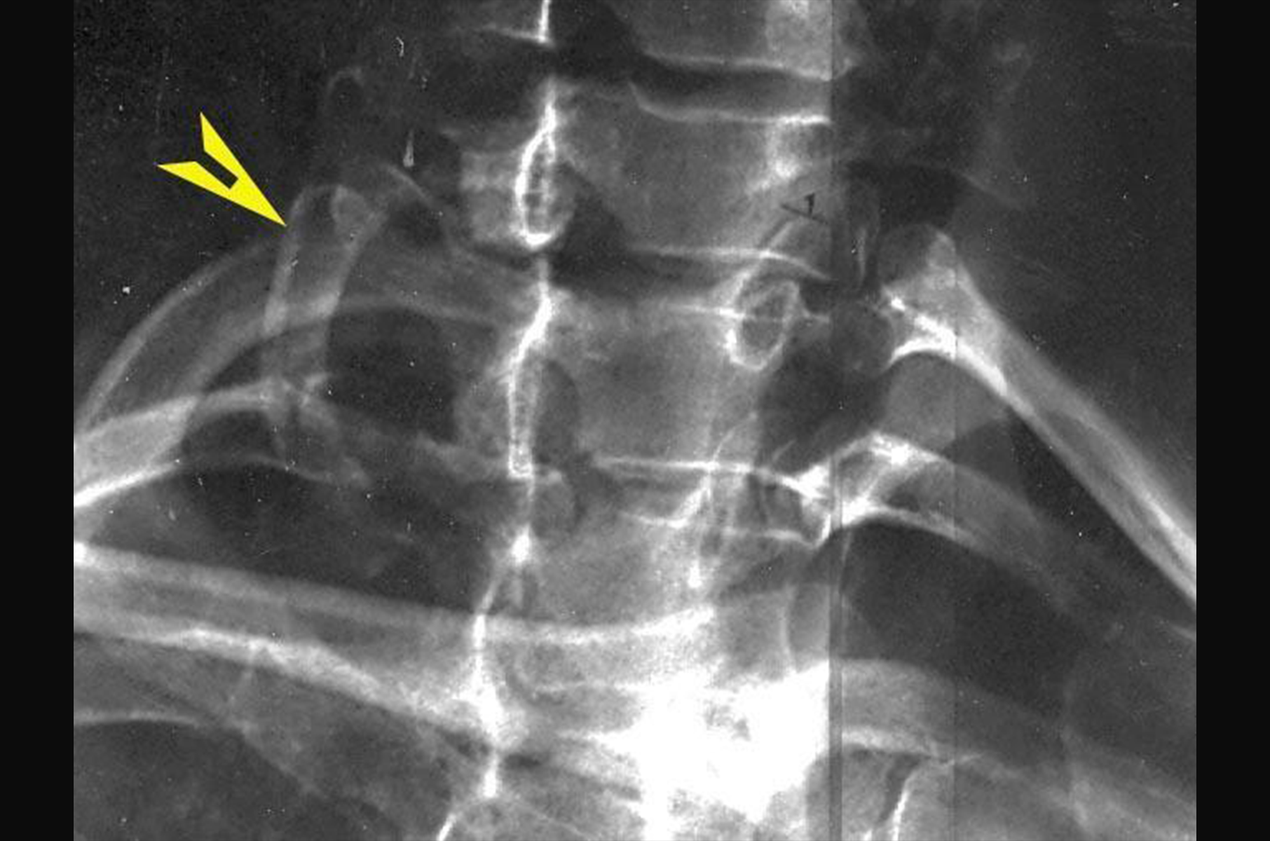
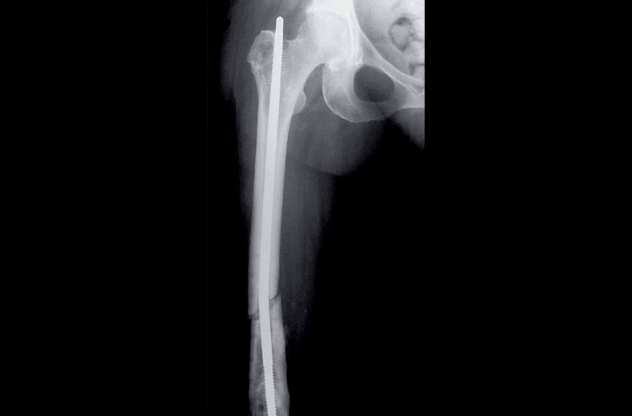
- X-rays 4-5: Identify the structure at the arrow.
Part 2: Clinical decision rules
Several medical professionals use set criteria when ordering radiographs. Compare and contrast the Pittsburgh and Ottawa rules for ordering radiographs. Which do you think is best? What populations are not covered by these rules? How could the physical therapy profession benefit from a standard set of rules?
Knowledge check
Each module will include a knowledge check section. This quiz is to check your knowledge on the topic from this week and it will also give you some idea of the types of questions asked on the graded quizzes. Click below to complete the knowledge check.


